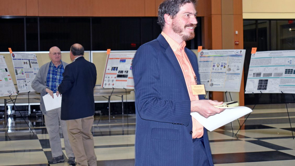
GREENVILLE, South Carolina – Organizers of the Wallace R. Roy Functional Radiology Symposium, which was held March 16 at Prisma Health, said they hope the event will foster increased collaboration between the hospital and local universities, including Clemson. Their long-term goal was to bring cutting-edge medical imaging research to the Upstate.
The symposium drew around 100 health care professionals, researchers and students, who came to learn how radiology can be used to do more than view bones and soft tissue. Functional radiology – also known as functional imaging – focuses on physiological changes that can be detected or measured, often by using tracers or sensors.

Jeff Anker, the Wallace R. Roy Distinguished Associate Professor in analytical chemistry who helped organize the symposium, is working to develop applications that will use conventional techniques to help improve surgical patients’ recoveries by minimizing the risk of post-operative infection.
Through his research, Anker said he is trying to get new information about what’s happening in the body during diseases, both chemically and mechanically.
“I’ve been making sensors that look at local acidity on an implanted medical device (such as a metal bone plate) in hopes of being able to detect infection early, monitor it during treatment to ensure eradication or learn that the treatment is not working so additional surgery is necessary, and to really understand the chemical environment so we can develop antimicrobial techniques and avoid infection in the first place,” Anker said.
Anker noted another use of functional radiology – functional MRIs that may help researchers unlock some of the secrets of the brain.
“If I am trying to look at the brain, I can see the anatomy beautifully with a conventional MRI,” Anker explained. “But if I want to see which parts of the brain light up, then I need to start making the MRI functional, meaning looking at the blood flow, or looking at some other signal. You look at the blood flow in the brain in order to see what areas of the brain light up. It’s very useful in understanding fundamental things, like memory, but also things like stroke and autism and also Alzheimer’s.”
There were several talks about functional MRI at the meeting. Dr. Andrei Holodny, chair of neuroradiology at Sloan Kettering, described how functional MRI measurement of brain activity connected with language could be used to help physicians plan whether and how to remove brain tumors while avoiding language deficiencies.
Similarly, Chris Whitlow at Wake Forest explained how he used functional MRI to study how repeated collisions during football practice affected brain activity in children playing football.
A major theme was how to use computers to better present and analyze images. Dr. Chris Rorden, co-director of the McCausland Center for Brain Imaging at USC, explained the difficulty of mapping brain regions after a patient has a stroke, and described methods to use the undamaged side as a reference. Ge Wang, an Endowed Professor at Rensselaer Polytechnic Institute, described methods to improve images using computer learning programs. Dr. John Absher at Prisma Health is leading an initiative to analyze existing databases of patient images to lean about indicators of neurodegenerative diseases such as Alzheimer’s disease.
Other talks focused on cardiovascular imaging to detect and treat aneurysms, orthopedic and cancer imaging, and genetic testing for developmental diseases.

Researchers from top universities and medical facilities including Prisma Health, Clemson, Yale, Greenwood Genetic Center, Sloan Kettering, Vanderbilt, University of Alabama Birmingham, and Wake Forest gave presentations during the daylong symposium, which also included a panel discussion and a poster session with 24 posters.
Beyond exploring the topic of functional radiology, the symposium was designed to be another step in a growing collaboration between Clemson University and Prisma Health, according to Dr. Ron Cowley, a neuroradiologist who began practicing at GHS in 1981.
“I’ve seen all this opportunity around Greenville, with researchers at Clemson and at Furman and at Bob Jones University, and the hospital system,” Cowley said, “and it’s been my dream for at least 20 years to see all those people come together.”
Cowley said this goal is closer than ever to being achieved through the Clemson University School of Health Research (CUSHR), a multidisciplinary unit of Clemson that facilitates medical research and scholarship.
“CUSHR is an effort that has been going on for three or four years,” Cowley said. “I asked them if we could put together some kind of consortium to help the researchers at Clemson and the clinicians and researchers (at Prisma Health) somehow get together so that they could give us what we need, and we could somehow help them focus on what they can do.”
Anker agreed that there is a need for more collaboration in the health-care field between Clemson and Prisma Health, formerly the Greenville Health System.
“We really need to have more collaborations with the hospital,” he said. “In particular, it would be really helpful to have more imaging capabilities. The hospital has an interest in being able to do better imaging and do more research, and Clemson certainly has an interest in being able to collaborate with physicians and radiologists who are going to be working on real problems.”
The Wallace R. Roy Functional Radiology Symposium was presented by CUSHR, SC BioCRAFT, Clemson University’s chemistry department and Prisma Health. It is named in honor of Clemson graduate Wallace R. Roy.
Get in touch and we will connect you with the author or another expert.
Or email us at news@clemson.edu

