They can’t be seen with the naked eye, but microorganisms are all around us. While most are harmless and some are beneficial, others can make us sick.
September 17 is International Microorganism Day, marking the day in 1683 when Dutch scientist Antonie van Leeuwenhoek sent a letter detailing the first microscopic observation of a single-celled organism we now call bacteria. The letter described “living animalcules” that van Leeuwenhoek gathered from his own mouth and those of others.
To commemorate the day, we’re highlighting three Clemson University College of Science faculty members whose research involves microorganisms.

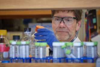
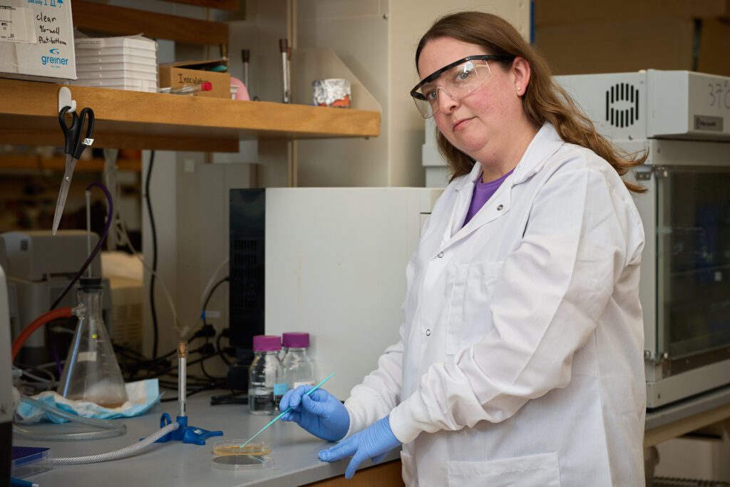
Emily Rosowski
Most humans inhale fungal spores daily. People with healthy immune systems can clear the spores without any problems, but those who are immunocompromised are susceptible to infection.
Emily Rosowski, an assistant professor in the Department of Biological Sciences, studies microbes with pathogenic potential in immunocompromised people. These people could have a genetic deficiency or take immunosuppressive drugs, such as transplant patients who take medication so their immune system does not attack and reject the new organ.
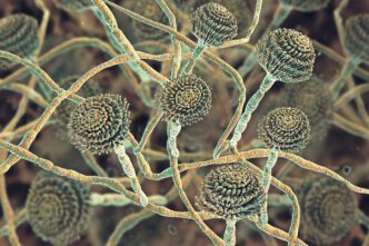
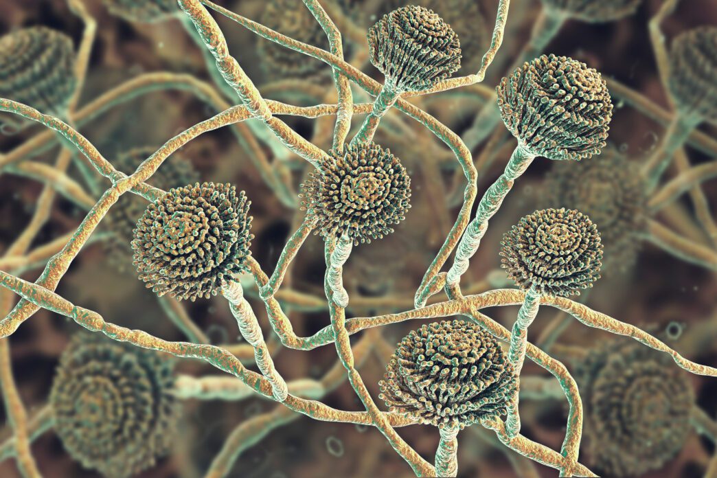
These types of infections are called opportunistic infections. Rosowski focuses explicitly on aspergillosis, the disease caused by the fungus Aspergillus fumigatus. Aspergillus fumigatus lives in our environment just about anywhere, mainly in the soil, and functions as a decomposer. The microorganism produces spores during its reproductive life cycle stage.
Rosowski’s research focuses on understanding the immune system’s first line of defense against these spores to determine what goes wrong in immunocompromised individuals. When people are infected, Rosowski says, “these small spores germinate into hyphae, which is a filamentous growth. That growth secretes enzymes that degrade your tissues so it can grow throughout your tissues and through all your body.”
There are antifungal drugs that can stem infection, but 50% of patients who take these drugs still die from the disease.
Rosowski uses zebrafish in the larval form to visualize via microscope what’s happening in the early stages of infection and how specific cells in the immune system react to the fungus. Rosowski and her team have found a particular receptor called Toll-like Receptor 2 that helps the immune system recognize and target the spores.
“The long-term goal is to identify the key pathways that your healthy innate immune system uses to control the fungus and then think about ways you could try to boost those pathways in immunosuppressed people,” Rosowski says.
Rosowski’s work has the potential to lead to a treatment that would prevent infections from taking hold in immunocompromised individuals.
Katelyn Walzer
Splash pads and pools are popular attractions in the summer as people seek to keep cool in the scorching heat. But that water can bring something other than relief from high temperatures.
Cryptosporidium is a parasitic microorganism that causes diarrheal disease and can linger in water for over a week. More surprisingly, it is resistant to chlorine.
Katelyn Walzer, an assistant professor in the Department of Biological Sciences, studies Cryptosporidium — specifically Cryptosporidium parvum, which infects cattle and humans. About 70% of 1- to 3-week-old calves are infected with Cryptosporidium. The infection usually spreads in cattle through fecal to oral transmission.
In humans, the parasite poses the most risks to malnourished children and the immunocompromised.
Historically, cryptosporidiosis has been an understudied disease. It is usually underreported since most instances do not require hospitalization. Most cases requiring medical intervention occur in developing countries that do not always have access to clean water.
The parasite can lead to malabsorption of nutrients, which stunts growth and leads to the inability to fully heal from the infection.
The parasite goes through a dormant form called an oocyst, forming a resilient wall that allows it to persist in water without a host. Humans need to ingest only 10 oocysts to become ill, but a single diarrheal episode could excrete millions.
When the parasite changes to its active form, it infects its host’s gut and goes through sexual reproduction. The process of the parasites’ becoming male or female is essential to its propagation in the host. Walzer is researching how the parasites switch from asexual to male or female by looking at which genes activate during different points of the parasite’s life cycle.
Walzer identified a transcription factor, Myb-M, that, when removed from Cryptosporidium, stops parasites from becoming male. When Myb-M is removed from Cryptosporidium-infected mice, the infection subsides.
“We can use sex in this parasite as a transmission blocking strategy and also as a continuous infection blocking strategy,” Walzer says.
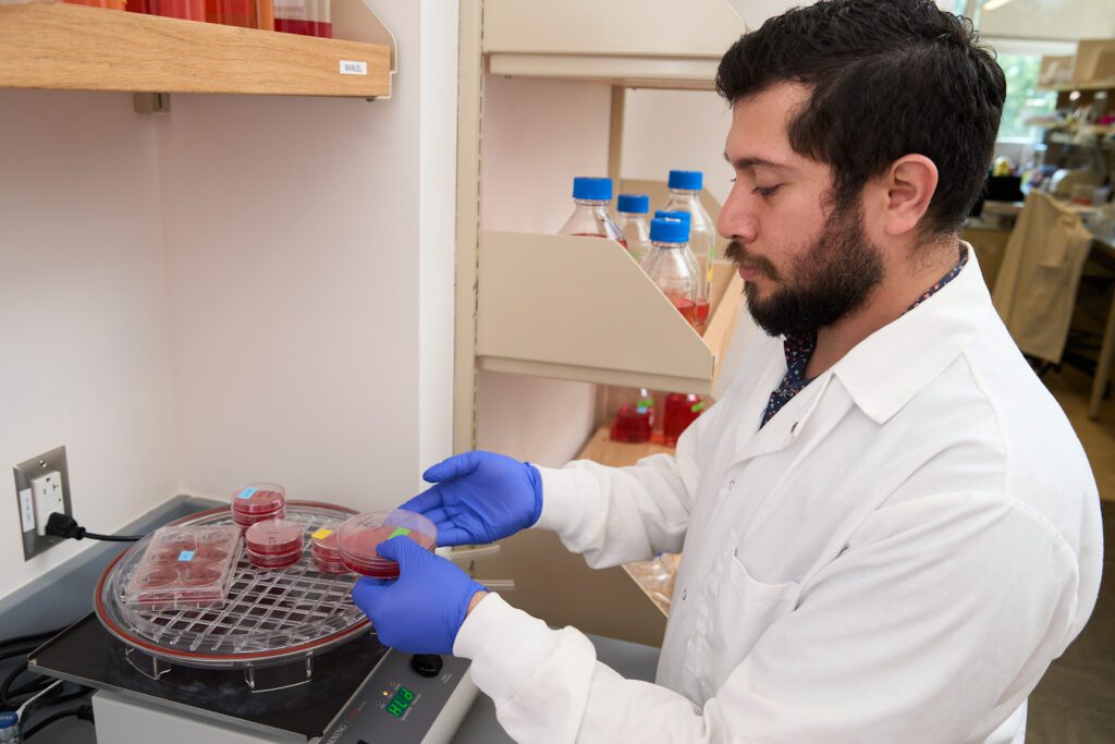
Manuel Fierro
Around two-thirds of the global population is at risk of contracting malaria because they live in areas where the disease can be transmitted by mosquitoes.
Health organizations estimate that malaria causes 600,000 deaths annually, mostly among children. Medicines exist but resistance against all antimalarials is reported globally, and the currently available vaccine provides only about 30% to 60% protection.
Manuel Fierro, an assistant professor in the Department of Genetics and Biochemistry, studies the cell biology of the microorganism that causes malaria. Malaria is caused by a single-cell eukaryotic parasite that invades other cells.
Fierro investigates the different biological pathways necessary for the parasite’s survival.
Malaria has developed resistance to every anti-malarial drug. With six different species of malaria that can infect humans, each geographic area responds differently to medicines.
“That’s why my type of research is important to continue increasing knowledge about malaria and which biological pathways or proteins are critical for its survival. That can then inform the rest of the community, and it could result in another much needed anti-malarial,” Fierro says.
Malaria infection and symptoms come from the microorganism invading the body and breaking red blood cells open. Fierro studies the active portion of the malaria life cycle, focusing on the deadliest species, Plasmodium falciparum. He uses human blood to culture the parasite and different molecular approaches to visualize proteins and discover their importance.
Malaria is a haploid organism, meaning it has one only copy of each gene in each cell. That means traditional knockout methods of removing one copy of genes are not viable. Fierro has instead developed processes to tune down the expression of a gene instead of entirely knocking it out.
To survive within the host’s body, malaria must remake the red blood cell it has invaded, programming it to bring nutrients to the parasite. It exports and sends hundreds of proteins to the red blood cell to modify the cell. While researchers have determined the key players involved in protein transport from malaria parasites to the red blood cell, there is a gap in knowledge about which compounds can hit these proteins and disable the parasite.
“Currently my group is trying to develop sensitive assays to measure protein export. The current methods we have to measure export take a long time, and our approach would be quicker and more reliable,” Fierro says. This assay could lead to large-scale screening to determine what compounds inhibit malaria’s ability to survive within a human host, which could have future treatment implications.